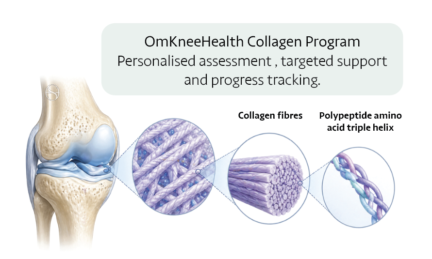
How each tissue is built
the collagen types that make up every part of the knee
Articular (chondral) cartilage
Collagen types present: II, IX, XI, VI, X
Articular cartilage is the smooth, white surface that caps the ends of the bones and lets them slide with almost no friction. About two-thirds of its dry weight is collagen, and the overwhelming majority of that (roughly 90 to 95 per cent) is type II. The type II fibrils do not work alone: they form a cross-linked co-polymer with types IX and XI, which regulate fibril size and lock the network together, while type VI forms the mesh right around each cartilage cell. Down at the bone, a thin band of calcified cartilage rich in type X anchors the whole layer to the skeleton, with small amounts of types III, XII and XIV present as well.
The way these fibres are arranged is as important as which type they are. In the surface zone the collagen runs parallel to the joint surface to resist shear; in the deep zone it turns to run vertically, anchoring the cartilage into bone; and in between the fibres curve through the middle in arches. Because cartilage has no blood supply and very slow cell turnover, this tissue heals poorly once it is damaged, which is why protecting it matters so much.
Meniscus
Collagen types present: I (outer), II (inner), VI
Each meniscus is a wedge of fibrocartilage, roughly seventy per cent water, with collagen making up about three-quarters of the dry weight. Its collagen changes from the outside in. The tough outer rim, the red zone, is around ninety per cent type I, a fibrous, tensile tissue built to be squeezed. The inner zone, the white zone, is different: it carries far more type II collagen (up to about sixty per cent there) with type I making up the rest, so it behaves more like articular cartilage, and it holds relatively more type VI around its cells.
The strong type I fibres run the long way around the C-shape. When the thigh bone presses down, the wedge is squeezed outward and these circumferential fibres pull tight to hold it, turning a downward squeeze into a spread-out load. This is hoop tension, and it is the meniscus’s single most important trick. Radial tie-fibres cross the circumferential ones like stitches to stop the tissue splitting. Because only the outer third has a blood supply, tears in the red zone can heal or be repaired, while tears in the avascular white zone rarely mend on their own.
Cruciate ligaments: the ACL and PCL
Collagen types present: I, III, II and X (entheses), IV, V, VI
The cruciate ligaments cross inside the knee and control rotation and front-to-back stability. They are dense, rope-like bands built from about ninety per cent type I collagen with roughly **ten per cent type III**, the finer form that gives a little flexibility and does much of the early work in healing. Their fibres have a wavy crimp that straightens under load, acting as a built-in shock absorber. Where the ligament meets bone, the tissue turns to fibrocartilage containing type II and type X; type IV appears in the small blood vessels, type VI acts as a gliding layer between fibre bundles, and type V helps set fibril size.
The anterior cruciate ligament sits inside the joint, bathed in joint fluid, with a patchy blood supply that is best near the thigh bone and poor near the shin. That environment is why a fully torn ACL often struggles to heal itself. During healing, or when a graft is used, the tissue first lays down weaker type III collagen and then remodels it toward strong, aligned type I over many months. The posterior cruciate ligament is thicker, better supplied and often heals well without surgery.
Collateral ligaments: the MCL and LCL
Collagen types present: I, III
The medial and lateral collateral ligaments run down the inside and outside of the knee and resist it being forced sideways. Like the cruciates they are dense type I collagen with a smaller amount of type III. The medial collateral ligament sits outside the joint capsule where blood reaches it easily, so it forms a stable clot and most injuries, even complete tears, heal well with time, bracing and guided rehabilitation. The lateral collateral ligament and the structures of the posterolateral corner heal less reliably and more often need a specialist’s attention.
Tendons: quadriceps, patellar and the iliotibial band
Collagen types present: I, III, V, II (entheses)
Tendons transmit the pull of muscle to bone. They are among the most type I collagen-rich tissues in the body, with roughly ninety-five per cent of their collagen being type I, laid down in tightly packed parallel bundles for pure tensile strength. A small amount of type III is present and rises during healing and with age, and type V helps regulate fibril diameter. Where a tendon inserts into bone it passes through a zone of fibrocartilage containing type II. Tendons respond best to steady, progressive loading rather than complete rest, and although a complete tear usually needs surgery, most tendon problems settle with graded rehabilitation.
Bone: the living scaffold
Collagen types present: I
Bone is often thought of as purely mineral, but its framework is collagen. About ninety per cent of bone’s organic matrix is type I collagen, and the mineral, a form of calcium phosphate, hardens onto that collagen scaffold. The collagen gives bone its toughness and resistance to cracking, while the mineral gives it stiffness and resistance to compression. Bone is constantly renewed, and load-bearing movement is part of what keeps that renewal healthy.
Think of collagen as the scaffold that holds your knee together. Your actions can influence your collagen health.
What happens to collagen when you hurt your knee
When you tear a ligament, a meniscus or a cartilage surface, what you are really tearing is collagen. The body’s first repair move is to lay down a quick, weaker type III scaffold, which it then remodels toward stronger, better-organised type I over the following months. Loading the tissue well, and at the right stage of healing, is what drives that remodelling.
Not every tissue rebuilds equally. Some, such as the collateral ligaments and the outer meniscus, recover well because they have a good blood supply. Others, such as the ACL and the inner cartilage, heal poorly on their own because of a poor blood supply and the joint environment they sit in. Knowing the difference tells you where good habits help most, and where you need clinical care.
Why collagen changes with age
With age, the body makes collagen more slowly and lays it down in a less organised way. Over time that reduces the resilience of cartilage, slows repair after injury, and can raise the risk of the next one. None of this is fixed. Collagen is a living tissue that responds to how you move, what you eat and how you recover, so there is plenty you can do to look after it.
Giving your collagen what it needs
- Vitamin C. Vitamin C contributes to normal collagen formation. It is found in citrus fruit, peppers and leafy greens.
- Protein. Provides the amino-acid building blocks the body uses to make and repair collagen.
- Collagen peptides. Studied alongside appropriate loading for joint and tendon health.
- Zinc and copper. Trace minerals involved in the cross-linking that gives collagen its structural strength.
- Sleep. Deep sleep is when much of the body’s repair and renewal happens. Protect it.
- Avoid smoking. Smoking impairs collagen and slows healing, working against everything else you do.
Movement matters. Collagen is not static. It remodels in response to the loads you put through it, which is why steady, progressive exercise strengthens these tissues and improves their resilience, while long spells of rest tend to weaken them.
Load your knee gradually and build up. Collagen rewards steady, progressive movement.
Keep exploring the Knee Passport
- Chondral surfaces: the smooth glide layer, in detail, and osteochondral health. → Read more
- The meniscus: your shock absorber, and how tears are managed. → Read more
- Ligaments of the knee: the cruciate and collateral stability system. → Read more
- Check your knee scores: validated self-checks to track your recovery. → Read more
- Rehabilitation protocols: structured, progressive loading for each stage. → Read more
- Clinic visits: assessment and a plan with the SportsHealing team. → Read more
References
- Ricard-Blum S. The collagen family. Cold Spring Harbor Perspectives in Biology, 2011.
- Eyre D. Collagen of articular cartilage. Arthritis Research, 2002.
- Fox AJ, Bedi A, Rodeo SA. The basic science of human knee menisci: structure, composition and function. Sports Health, 2012.
- Duthon VB and colleagues. Anatomy of the anterior cruciate ligament. Knee Surgery, Sports Traumatology, Arthroscopy, 2006.