THE MECHANICS OF MOTION
How the Knee Locks and Unlocks
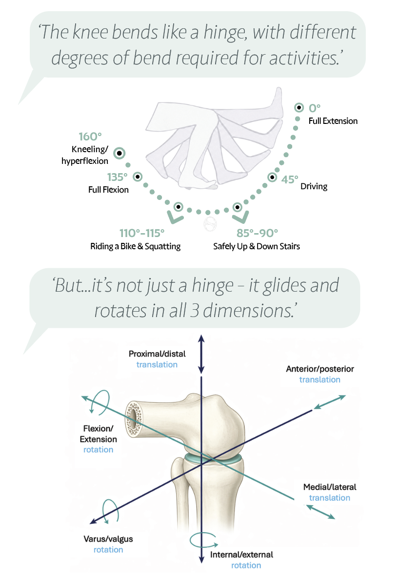
Roll, then glide. The knee does not bend like a simple door hinge. As you flex, the rounded end of the femur first rolls backwards on the tibia; only after roughly the first 25 degrees of bend does it begin to also glide. This roll-then-glide, together with the constantly shifting pivot, is what creates the room for the knee to bend deeply, beyond a right angle, without the bones colliding. The slight backward tilt of the top of the tibia, about 7 to 10 degrees, helps make that deep bend possible.
Locking straight, without effort. Straightening the knee has a clever finish. The smaller outer (lateral) side of the joint runs out of surface first, at around 30 degrees from straight, so the larger inner (medial) side keeps travelling through the last stretch. That mismatch forces the shin to rotate very slightly, wedging the bones, menisci and ligaments into a tight, stable position over the final few degrees. This is the screw-home mechanism, and the important point is that it needs no muscle at all: a fully straightened knee locks and holds itself, which is why standing upright feels so effortless.
The muscle that unlocks it. To bend again, the lock must first release. A small muscle tucked behind the knee, the popliteus, quietly rotates the shin the other way to unlock the joint just before bending begins. You never notice it working, but it is the switch that lets a locked, straight knee start to fold.
A pivot on the inside. The two sides of the knee do not move identically. The inner (medial) side glides more and acts as a relatively stable pivot, while the outer (lateral) side rolls and travels further. The result is a small, natural rotation built into every bend and straighten, the knee turning gently around its inner edge.
‘Think of straightening your knee fully as clicking it into a locked, restful position, and bending as gently unlocking it again. Trusting that lock is part of moving with confidence.’
TRAINING THE KNEE
Open Chain and Closed Chain — Why It Matters in Rehabilitation
One of the most useful ideas in knee rehabilitation is the difference between closed-chain and open-chain movement. It quietly shapes almost every exercise programme.
Closed chain means your foot is planted and you move your body over it — squats, lunges, step-ups, standing up from a chair, going up and down stairs. The whole leg works as a unit: hip, knee and ankle share the job, the muscles around the knee tighten together, and the joint is well supported from every direction. Because the load is shared and the muscles stabilise the joint as a team, closed-chain movements are the most like real life, and are often where rehabilitation begins.
Open chain means your foot is free and you move just the lower leg — for example straightening the knee against resistance while seated (a leg extension), or a seated hamstring curl. These isolate one muscle group at a time, which makes them excellent for rebuilding a specific weak link, but they concentrate the load on one part of the knee rather than spreading it.
Why the difference matters. Different exercises stress different structures. A resisted open-chain leg extension draws the shin forwards and asks more of the ACL, and it loads the kneecap most as the knee nears straight. A closed-chain squat, by contrast, loads the kneecap and cartilage more as the knee bends deeper, and the deeper and heavier you go, the more the joint has to handle. Neither type is simply “good” or “bad” — each has its place.
This is exactly why, after an ACL injury or with kneecap pain, a physiotherapist chooses the type, range and depth of each exercise deliberately: often favouring closed-chain work early because it is stable and functional, and introducing open-chain work to target a specific muscle once the knee is ready. Load is added gradually and on purpose, so that strength is built step by step rather than all at once.
Understanding why your programme looks the way it does — why some exercises come first, why depth and resistance are added slowly — turns rehabilitation from a list of instructions into something you can trust and take ownership of.
WHAT AFFECTS MOVEMENT QUALITY
The Factors That Control How Your Knee Moves
ACTIVE CONTROL
- Quadriceps — primary knee extensor and stabiliser
- Hamstrings — flex the knee and protect the ACL
- Hip abductors and glutes — control knee alignment
- Calf muscles — control foot and ankle position
- Nervous system — coordinates timing and force
PASSIVE RESTRAINTS
- ACL, PCL — sagittal plane stability
- MCL, LCL — coronal plane stability
- Posterolateral corner — rotational stability
- Menisci — load distribution and proprioception
- Joint capsule — limits end-range movement
One link in a chain. The knee never works alone. It sits in the middle of a chain that runs from the spine through the hip and down to the ankle, and the segments above and below strongly influence it. Weakness or stiffness at the hip or ankle changes the demands on the knee, which is exactly why good rehabilitation looks beyond the knee itself to the glutes, hips and calves.
Control can be trained. Reassuringly, the knee’s stability is not fixed by anatomy alone. Coordinated muscle timing and reflexes can compensate for a great deal, and targeted neuromuscular training has been shown to help the joint move with more stability and confidence, even where the passive structures are doing less of the work.
YOUR SELF-CHECK: After sitting for an hour, stand up and take 10 slow steps. Does your knee improve after those first steps? That warming-up process is the synovial fluid distributing through the joint. If it does not improve, speak to your clinician.
REFERENCES — CLINICAL REFERENCES
- Dye SF (1996) — The knee as a biologic transmission with an envelope of function: a theory. *Clinical Orthopaedics and Related Research* 325:10–18.
- Hallén LG, Lindahl O (1966) — The “screw-home” movement in the knee-joint. *Acta Orthopaedica Scandinavica* 37(1):97–106.
- Reynolds RJ, Michelet A, Müller JH, Saffarini M (2022) — Kinematics of the Native Knee. In: Becker, Hirschmann & Kort (eds.), *Basics in Primary Knee Arthroplasty*. Springer.
- Hazari A, Maiya AG, Nagda TV (2021) — Kinematics and Kinetics of the Knee Joint. In: *Conceptual Biomechanics and Kinesiology*. Springer. Source for the locking (screw-home) mechanism, the roll-to-glide transition, the popliteus as the unlocking muscle, and open- versus closed-chain kinetics.